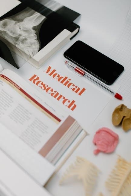
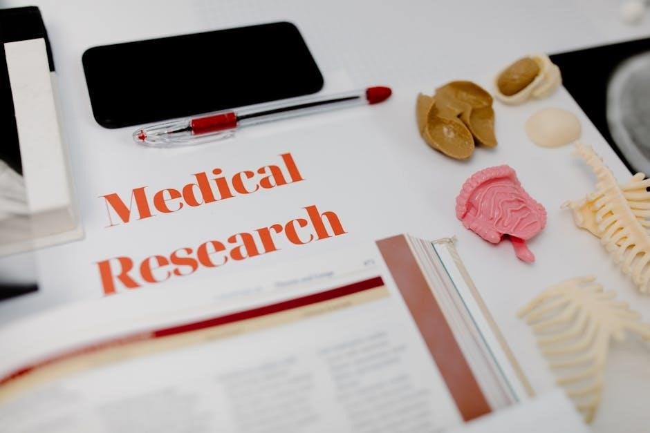
Welcome to the Anatomy & Physiology laboratory manual‚ designed to guide students through hands-on exploration of human structure and function․ This manual provides structured exercises‚ practical applications‚ and critical thinking challenges to enhance learning․ Each lab activity aligns with key concepts‚ ensuring a comprehensive understanding of anatomical and physiological principles․
1․1 Purpose and Importance of the Laboratory Manual
The laboratory manual serves as a vital resource for hands-on exploration of anatomical structures and physiological processes․ It bridges theoretical knowledge with practical application‚ enhancing critical thinking and observational skills․ By engaging in structured exercises‚ students develop a deeper understanding of complex concepts‚ preparing them for real-world applications in healthcare and scientific fields․
1․2 Essential Laboratory Tools and Equipment
The laboratory manual requires essential tools such as microscopes‚ dissection instruments‚ and measurement devices․ These tools enable students to explore anatomical structures‚ conduct experiments‚ and analyze physiological processes․ Proper use of equipment ensures accurate observations and safe lab practices‚ fostering a hands-on learning environment for anatomy and physiology studies․
1․3 Safety Precautions and Laboratory Etiquette
Adhering to safety protocols is crucial in the lab․ Wear protective gear like goggles and gloves‚ and handle chemicals with care․ Follow proper disposal procedures and know emergency exit routes․ Respect shared spaces‚ clean up after use‚ and avoid distractions to maintain a safe and efficient learning environment for all students․

Organization of the Body and Anatomical Terminology
This section introduces the hierarchical structure of the human body‚ from cells to systems‚ and explores anatomical terminology‚ enabling precise communication about body regions‚ directions‚ and positions․
2․1 Levels of Organization in the Human Body
The human body is organized into a hierarchical structure‚ beginning with cells‚ which form tissues‚ then organs‚ and finally organ systems․ Each level builds on the previous one‚ enabling complex functions․ This organization is essential for understanding how the body maintains homeostasis and overall physiological balance․
2․2 Anatomical Planes and Directions
Anatomical planes divide the body into sections: sagittal (vertical)‚ frontal (coronal‚ vertical)‚ and transverse (horizontal)․ Directions include anterior (front)‚ posterior (back)‚ dorsal (upper back)‚ ventral (belly)‚ proximal (near origin)‚ and distal (far from origin)․ These terms are essential for precise anatomical descriptions and lab dissections․
2․3 Body Cavities and Membranes
Body cavities‚ such as thoracic‚ abdominal‚ and pelvic‚ house internal organs․ Membranes like pleura‚ pericardium‚ and peritoneum line these cavities‚ reducing friction and protecting organs․ Understanding these structures aids in lab dissections and grasping anatomical relationships․ The study of body cavities and membranes is crucial for identifying organ locations and spatial orientations․
Microscopy and Histology
Explore microscopic structures using compound and electron microscopes․ Learn tissue preparation‚ staining techniques‚ and identification of cellular structures․ Essential for understanding histological features in anatomy and physiology labs․
3․1 Types and Proper Use of Microscopes
Learn about compound and electron microscopes‚ their components‚ and proper handling techniques․ Understand how to prepare slides‚ focus specimens‚ and maintain instruments for optimal viewing․ Proper microscopy skills are essential for analyzing histological samples in anatomy and physiology labs․
3․2 Preparing and Staining Tissue Samples
Master the steps of tissue preparation‚ including fixation‚ sectioning‚ and staining․ Fixation preserves tissue structure‚ while staining enhances visibility under a microscope․ Proper techniques ensure accurate histological observations․ Always follow safety protocols when handling chemicals and biological samples to maintain a safe laboratory environment․
3․3 Identifying Basic Tissue Types Under the Microscope
Learn to distinguish epithelial‚ connective‚ muscle‚ and nervous tissues under the microscope․ Observe unique cellular structures and arrangements․ Proper staining techniques enhance visibility‚ aiding in accurate identification․ Practice identifying histological features to build a strong foundation in tissue recognition and classification․
The Integumentary System
Explore the structure and function of the skin‚ the body’s largest organ‚ and its accessory organs like hair‚ nails‚ and glands․ Examine histological samples and sensory receptors through lab activities to understand protective‚ regulatory‚ and sensory roles in maintaining homeostasis․
4․1 Structure and Function of the Skin
The skin‚ composed of the epidermis‚ dermis‚ and hypodermis‚ serves as a protective barrier‚ regulates body temperature‚ and aids in sensation․ Lab activities include histological examinations of skin layers and identifying sensory receptors‚ emphasizing the skin’s role in maintaining homeostasis and overall health through its complex functions and structures․
4․2 Lab Activities: Skin Histology and Sensory Receptors
Lab activities focus on examining skin histology using microscopes to identify layers like the epidermis‚ dermis‚ and hypodermis․ Students analyze sensory receptors‚ such as Meissner’s and Pacinian corpuscles‚ to understand their roles in sensation․ Practical exercises include preparing histological slides and correlating skin structures with their functional roles in protection and sensory perception․
4․3 Common Integumentary System Disorders
This section explores prevalent integumentary system disorders‚ including skin cancer‚ eczema‚ psoriasis‚ and fungal infections․ Emphasis is placed on understanding their causes‚ symptoms‚ and effects on skin integrity and sensory function․ Lab activities include clinical case studies to identify and manage these conditions‚ enhancing diagnostic and therapeutic skills․
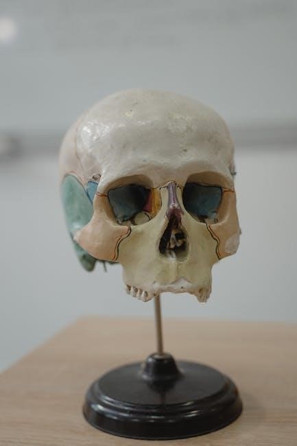
The Skeletal System
The skeletal system provides structural support‚ protects vital organs‚ and facilitates movement․ It consists of bones‚ joints‚ and cartilage‚ working together to maintain posture and enable mobility․
5․1 Classification and Structure of Bones
Bones are classified as long‚ short‚ flat‚ irregular‚ or sesamoid based on shape and function․ Their structure includes compact and spongy bone tissue‚ periosteum‚ and endosteum․ Long bones like the femur have a diaphysis and epiphyses‚ while flat bones such as the skull protect internal organs․ Each type adapts to specific roles․
5․2 Joints and Movement: Types and Functions
Joints are classified as synarthroses‚ amphiarthroses‚ or diarthroses based on movement․ Synarthroses are immovable‚ like skull sutures․ Amphiarthroses allow limited movement‚ such as intervertebral discs․ Diarthroses are freely moving‚ like the shoulder․ Each joint type facilitates specific ranges of motion‚ enabling functional movements essential for daily activities‚ as explored in lab exercises․
5․3 Lab Activities: Bone and Joint Dissection
Lab exercises involve dissecting bones and joints to explore their structure and function․ Students identify bone markings‚ joint types‚ and ligament attachments․ Activities include analyzing synovial joints‚ observing joint movements‚ and understanding the role of cartilage and synovial fluid․ Practical dissection enhances comprehension of skeletal morphology and articulations․
The Muscular System
The muscular system enables movement‚ supports posture‚ and facilitates body functions․ This section explores skeletal‚ smooth‚ and cardiac muscles‚ focusing on their structure‚ function‚ and physiological roles․
6․1 Types of Muscles and Their Functions
The muscular system comprises three types: skeletal‚ smooth‚ and cardiac muscles․ Skeletal muscles enable voluntary movement‚ smooth muscles regulate involuntary actions like digestion‚ and cardiac muscles pump blood․ Each type has distinct structures and functions‚ essential for movement‚ stability‚ and internal processes․
6․2 Muscle Physiology and Contraction Mechanisms
Muscle contraction occurs due to the sliding filament theory‚ where actin and myosin fibers interact․ This process is initiated by nerve stimulation‚ triggering calcium release‚ which binds to troponin and tropomyosin‚ uncovering myosin binding sites․
Energy from ATP drives the cross-bridge cycle‚ enabling movement and contraction‚ essential for bodily motion and function․
6;3 Lab Activities: Muscle Dissection and Physiology Experiments
Lab activities include dissecting skeletal muscles to identify structural components and exploring physiological mechanisms․ Students conduct experiments to measure contraction force and observe nerve stimulation effects․ These hands-on exercises reinforce understanding of muscle function‚ enabling practical application of theoretical concepts like the sliding filament theory and cross-bridge cycle․
The Nervous System
The nervous system controls and coordinates body functions‚ enabling sensory perception‚ motor responses‚ and cognitive processes․ It comprises the central and peripheral nervous systems‚ working together to maintain homeostasis and facilitate complex bodily functions․
7․1 Structure and Function of Neurons
Neurons are specialized cells designed to transmit information through electrical and chemical signals․ They consist of a cell body‚ dendrites‚ and an axon․ Dendrites receive signals‚ while the axon transmits them to other neurons or effector cells‚ enabling communication and coordination within the nervous system․
7․2 Reflexes and Nerve Impulse Transmission
Neurons transmit signals through action potentials‚ generating electrical impulses that travel along axons․ Reflexes involve rapid‚ involuntary responses to stimuli‚ requiring integration of sensory and motor neurons․ Synaptic transmission allows signals to pass between neurons‚ enabling coordinated physiological responses and maintaining homeostasis․
7․3 Lab Activities: Nerve Dissection and Reflex Testing
Lab activities include nerve dissection to observe structural components and reflex testing to demonstrate neural pathways․ Students analyze nerve tissue under microscopes and conduct tests to assess reflex responses‚ ensuring proper safety protocols and equipment use․ These exercises enhance understanding of neural communication and reflex mechanisms․
The Digestive System
The digestive system is explored through its anatomy‚ physiology‚ and lab activities․ Students examine the digestive tract‚ enzyme functions‚ and absorption processes‚ enhancing their understanding of digestion and nutrient uptake․
8․1 Anatomy of the Digestive Tract
The digestive tract includes the mouth‚ esophagus‚ stomach‚ small intestine‚ and large intestine․ Each region has specialized structures‚ such as villi in the small intestine‚ to facilitate digestion and absorption․ Lab activities involve identifying these structures and understanding their roles in breaking down food into nutrients for the body․
8․2 Physiology of Digestion and Absorption
Digestion involves the enzymatic breakdown of food into smaller molecules‚ while absorption transfers nutrients into the bloodstream․ The small intestine uses villi to increase surface area‚ enhancing nutrient uptake․ Nervous and hormonal regulation ensures efficient digestion and absorption‚ maintaining energy and nutrient balance for bodily functions․ Lab experiments demonstrate these processes․
8․3 Lab Activities: Digestive Enzyme Experiments
Lab activities focus on exploring digestive enzyme function through controlled experiments․ Students analyze how enzymes like amylase and pepsin break down substrates under varying conditions‚ such as pH levels․ Practical exercises include testing enzyme activity in simulated stomach and intestinal environments‚ demonstrating the role of enzymes in digestion and nutrient absorption processes․

The Circulatory System
The circulatory system‚ comprising the heart‚ blood vessels‚ and blood‚ transports oxygen‚ nutrients‚ and hormones․ Lab activities include heart dissection and blood pressure measurement experiments․
9․1 Structure and Function of the Heart
The heart is a muscular‚ four-chambered organ with atria and ventricles․ Valves regulate blood flow‚ and septa separate oxygenated and deoxygenated blood․ Lab activities include dissecting the heart to explore its internal structures and their roles in circulation․
9․2 Blood Vessels and Blood Flow
Blood vessels include arteries‚ veins‚ and capillaries‚ each with distinct roles in transporting blood․ Arteries carry oxygenated blood away from the heart‚ while veins return deoxygenated blood․ Capillaries enable nutrient and waste exchange․ Lab activities involve studying vessel histology and measuring blood flow dynamics․
9․3 Lab Activities: Heart Dissection and Blood Pressure Measurement
Lab activities include heart dissection to explore cardiac structure and blood pressure measurement to analyze circulatory function․ These exercises provide hands-on experience‚ enhancing understanding of cardiovascular physiology and the regulation of blood flow․ Students gain practical skills while visualizing key anatomical features and physiological processes․
The Respiratory System
The respiratory system is essential for exchanging oxygen and carbon dioxide through breathing․ This section explores its structure‚ function‚ and related lab activities to enhance understanding effectively․
10․1 Anatomy of the Respiratory Tract
The respiratory tract begins with the nasal cavity and mouth‚ leading through the pharynx‚ larynx‚ and trachea․ It branches into bronchi‚ bronchioles‚ and ultimately alveoli‚ facilitating gas exchange․ This structured pathway ensures efficient airflow and oxygenation of blood‚ forming the foundation of respiratory function and gas exchange mechanisms in the human body․
10․2 Physiology of Breathing and Gas Exchange
Breathing involves the inhalation of oxygen and exhalation of carbon dioxide‚ regulated by the diaphragm and intercostal muscles․ Gas exchange occurs in alveoli‚ where oxygen diffuses into blood and carbon dioxide into lungs․ This essential process maintains oxygenation of tissues and removal of metabolic waste‚ critical for cellular respiration and overall bodily function․
10․3 Lab Activities: Lung Capacity and Gas Exchange Experiments
Lab activities include spirometry to measure lung capacities and simulate gas exchange․ Students explore vital capacity‚ tidal volume‚ and alveolar ventilation․ Experiments using varying oxygen levels demonstrate gas diffusion principles‚ enhancing understanding of respiratory physiology and its role in maintaining homeostasis through practical‚ hands-on experiences․
Review and Assessment
This section summarizes key concepts‚ addresses common lab mistakes‚ and provides guidelines for final assessments and lab reports‚ ensuring comprehensive understanding and preparedness․
11․1 Key Concepts and Terms
This section reviews essential anatomical and physiological terminology‚ emphasizing critical concepts such as anatomical planes‚ tissue types‚ and physiological processes․ Key terms are defined to reinforce understanding of body systems and laboratory techniques‚ ensuring mastery of foundational principles․
11․2 Common Lab Mistakes and Troubleshooting
Common lab errors include improper use of equipment‚ incorrect tissue identification‚ and miscalculations during experiments․ Troubleshooting involves reviewing procedures‚ seeking instructor guidance‚ and practicing techniques․ Attention to detail and adherence to protocols are crucial for accurate results and safe laboratory practices․
11․3 Final Assessment and Lab Report Guidelines
The final assessment evaluates students’ understanding of anatomical and physiological concepts through lab reports and practical exams․ Ensure accuracy in data collection and clear presentation of results․
Lab reports should include objectives‚ materials‚ procedures‚ results‚ and conclusions․ Use proper scientific terminology and maintain organization for clarity․ Proofread for errors to ensure professional quality․